Main » Scientific articles » Artificial Intelligence in TMJ Visualization: 5 Studies and the Transformation of Clinical Diagnostics
Artificial Intelligence in TMJ Visualization: 5 Studies and the Transformation of Clinical Diagnostics
In the era of rapid digitalization contemporary dentistry is developing as one of the most dynamic fields of medical practice, actively integrating digital technologies and interdisciplinary approaches.
Artificial intelligence and modern digital platforms are becoming key components of the transformation of imaging-diagnostic processes in temporomandibular joint pathology.
Artificial intelligence as a key tool
The systematic review included five studies in which CBCT, MRI and panoramic radiography were used, demonstrating diagnostic accuracy ranging from moderate to high depending on the target task and study methodology.
The authors note that the current literature reflects a profound transformation of approaches to TMJ imaging — from manual interpretation of images to hybrid workflows, where AI provides increased reproducibility, detection of early osteoarthritic and morphological changes, as well as support for clinical decision-making; at the same time, standardization of model training protocols and external validation on transnational patient samples are critically important.
Diagnostic ecosystem: structure and components
Within the review the role of multimodal approaches is traced, where CBCT and MRI data complement clinical examination information and anamestic data for comprehensive assessment of the TMJ.
CBCT: identification of degenerative and morphological changes
CBCT combined with machine learning algorithms was most often used to navigate the joint anatomy and detect structural changes, such as erosions of articular surfaces, subchondral sclerosis, osteophytes and deformities of the condylar region; in clinical practice this opens the possibility of earlier identification of patients with progressive osteoarthritis and planning corrective interventions.
MRI and panoramic radiography: functions and limitations
MRI remains the gold standard for assessment of the soft tissue components of the TMJ — the disc, the retrodiscal space, synovial fluid; AI-based MRI methods are capable of increasing sensitivity to internal derangements, however limitations are the quality and standardization of scanning protocols. Panoramic radiography provides limited useful information for structural analysis of the TMJ, AI applications on panoramic images are so far less informative for early diagnosis of intra-articular changes.
Validation, standardization and clinical integration
Key barriers to the implementation of AI in clinical practice remain small sample sizes, lack of external validation and variability of imaging protocols; to overcome these limitations multicenter studies, synchronization of clinico-radiological protocols and inclusion of models within existing diagnostic criteria are necessary.
The authors emphasize the importance of explainable AI solutions, which increase the transparency of algorithmic conclusions and can reduce clinicians’ excessive dependence on «black box», while simultaneously requiring the development of regulatory and ethical standards for use in dental practice.
Pune as a strategic platform
The study from Pune illustrates the transnational interest in the topic, but also demonstrates typical limitations — small patient cohorts and lack of external validation of models; this underscores the need to expand samples, involve multidisciplinary teams and share data to increase the clinical applicability of the results.
Interdisciplinary cooperation between radiologists, maxillofacial surgeons, clinical dentists and data specialists is a prerequisite for creating reproducible and clinically relevant models that can be integrated into clinic workflows.
Conclusion and expert commentary
AI-assisted TMJ imaging demonstrates the potential to increase diagnostic accuracy and reproducibility of interpretations, especially in the detection of early degenerative changes; however, to move from research evidence to everyday clinical practice standardized scanning protocols, external validation of models on large and diverse samples, assessment of the impact of AI on clinical outcomes and development of explainable interfaces for clinicians are necessary.
Practical recommendations for implementation: formation of multicenter cohorts and shared databases, standardization of CBCT and MRI parameters for the TMJ, inclusion of AI assessments in clinical protocols in pilot mode with outcome monitoring, emphasis on algorithm explainability and educational initiatives to increase clinicians’ digital literacy.
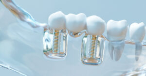
In modern implantology, based on digital technologies and interdisciplinary clinical practice, the problem of chronic inflammation around implants, caused by
We use cookies to improve website performance, analyze traffic, and personalize content. You can accept all cookies or customize their use. Learn more in our Cookie Policy.
Functional
Always active
Technical storage or access is strictly necessary for the legitimate purpose of making possible the use of a particular service expressly requested by the subscriber or user, or solely for the purpose of carrying out the transmission of a message over an electronic communications network.
Preferences
The technical storage or access is necessary for the legitimate purpose of storing preferences that are not requested by the subscriber or user.
Statistics
Technical storage or access used solely for statistical purposes.The technical storage or access that is used exclusively for anonymous statistical purposes. Without a subpoena, voluntary compliance on the part of your Internet Service Provider, or additional records from a third party, information stored or retrieved for this purpose alone cannot usually be used to identify you.
Marketing
Technical storage or access is necessary to create user profiles for the purpose of sending advertising or to track a user across a website or multiple websites for similar marketing purposes.