
Guided Endodontic Access: experience of more than 200 practitioners and the formation of a paradigm of precise endodontics
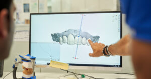
Contemporary dentistry is rapidly transforming under the influence of digital technologies and interdisciplinary approaches, altering clinical and educational practices worldwide.