Main » Uncategorized » Guided Endodontic Access: experience of more than 200 practitioners and the formation of a paradigm of precise endodontics
Guided Endodontic Access: experience of more than 200 practitioners and the formation of a paradigm of precise endodontics
Contemporary dentistry is rapidly transforming under the influence of digital technologies and interdisciplinary approaches, altering clinical and educational practices worldwide.
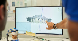
Guided endodontic access (GEA) is regarded as an integrated protocol combining CBCT, intraoral scanning, digital planning and 3D‑printed guides, which demonstrates clinical advantages when working with obliterated root canals and complex anatomical configurations.
Technology as a key tool
Guided endodontic access is not simply a technological novelty — it is an element of high‑precision navigation, providing reproducibility and predictability of access to the canals, especially in cases of complete or partial lumen obliteration. Key components of the protocol include high‑quality tomography with adequate voxel resolution, accurate registration of CBCT data and the intraoral scan, digital modeling of the access trajectory taking into account the thickness of dentin walls and the coronal neck, as well as fabrication of the guide with controlled tolerance of the seating recess and guiding sleeve.
Clinical mechanics and limitations
In planning, possible sources of error must be taken into account — inaccurate alignment of DICOM and STL files, micro‑mobility of the template during drilling, angular deviation of the drill trajectory and variability of dentin tissue density; critical parameters are the thickness of the cortical plate, distance to perforating structures and the length of the predetermined access. To minimize risks it is recommended to verify the fit of the guide intraorally prior to drilling, to use guiding sleeves compatible with the drill system, and to practice the protocol on models or simulators before moving to clinical cases.
Educational ecosystem: structure and content
A cross‑sectional study covering more than 200 practicing dentists revealed high awareness and a positive attitude toward GEA; the main barriers to adoption were identified as funding and limited access to clinically validated training. Training should go beyond software demonstrations — it is necessary to develop competencies in image segmentation, digital trajectory planning, guide design and quality control of 3D‑printing, combined with clinical training on phantoms and under the supervision of experienced mentors.
Recommendations for integration into the curriculum
Modules on navigation and 3D design should be included in continuous professional development and postgraduate programs; emphasis should be placed on interdisciplinary cooperation — endodontics, prosthetics, digital laboratories and radiology — to ensure reproducibility of clinical outcomes. Competency assessment should be based on objective metrics — accuracy of guide positioning, percentage of successful canal exposure without perforation, procedure time — and be accompanied by feedback and documentation of results for subsequent validation of protocols.
Ha’il as a strategic platform
A local study in Ha’il confirms transnational trends: specialists with higher academic status and greater clinical experience demonstrate greater readiness to integrate GEA, which underscores the role of professional networks and continuing education programs in accelerating adoption. For the region, it is strategically important to develop reproducible training models and infrastructure — simulation centers, access to 3D‑printing laboratories and mentorship programs — that reduce entry barriers and improve the quality of care.
Conclusions and clinical recommendations
Guided endodontic access demonstrates that modern endodontics is evolving toward an integrated digital ecosystem based on knowledge sharing and clinical cooperation. For practicing clinicians it is advisable to implement GEA gradually — start with carefully selected cases of obliteration, ensure standardized planning and quality control of guide production, document outcomes and complications, and participate in continuing education programs. For educational institutions it is recommended to integrate into curricula modules on CBCT‑segmentation, digital planning, 3D‑design and protocol validation, as well as to develop interdisciplinary practical laboratories and mechanisms for conducting multicenter studies to confirm the long‑term effectiveness of the method.
Dental technicians and the digital future: why adaptation secures the profession Introduction Julia Glancey, a dental technician and commentator, published
Case report: Bleach‑shade full‑smile rehabilitation using KATANA Zirconia YML and CERABIEN ZR Introduction / Background Authors Kostia Vyshamirski and Dr
We use cookies to improve website performance, analyze traffic, and personalize content. You can accept all cookies or customize their use. Learn more in our Cookie Policy.
Functional
Always active
Technical storage or access is strictly necessary for the legitimate purpose of making possible the use of a particular service expressly requested by the subscriber or user, or solely for the purpose of carrying out the transmission of a message over an electronic communications network.
Preferences
The technical storage or access is necessary for the legitimate purpose of storing preferences that are not requested by the subscriber or user.
Statistics
Technical storage or access used solely for statistical purposes.The technical storage or access that is used exclusively for anonymous statistical purposes. Without a subpoena, voluntary compliance on the part of your Internet Service Provider, or additional records from a third party, information stored or retrieved for this purpose alone cannot usually be used to identify you.
Marketing
Technical storage or access is necessary to create user profiles for the purpose of sending advertising or to track a user across a website or multiple websites for similar marketing purposes.