Main » Uncategorized » Blended Dentistry: transnational standardization of workflows and transformation of clinical and laboratory practice
Blended Dentistry: transnational standardization of workflows and transformation of clinical and laboratory practice
Digital dentistry forms a new paradigm of clinical and laboratory practice, in which the integration of digital tools and materials generates transnational ecosystems of care based on standardization and validation.
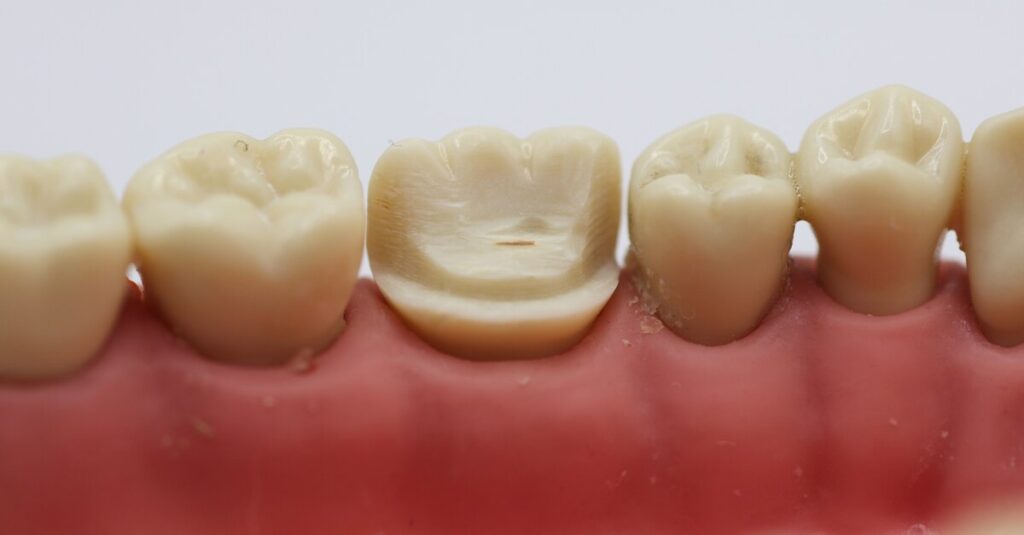
Blended dentistry is regarded as a workflow combining the classical principles of preparation for dentin pins and modern digital methods of designing and manufacturing indirect restorations — in particular, the fabrication of zinc-oxide and zirconia pinlay restorations, with sequential verification by thermography, digital colorimetry, intraoral scanning and milling in laboratory cooperation.
Technology as a key factor
Digital scanning and milling function not only as means of recording geometry but also as components of a reproducible workflow chain that requires synchronization of clinical and laboratory standards; such components are: intraoral scanners with high image coherence, CAD/CAM platforms for design and industrial milling stations operating with materials like multilayer zirconia Alien Multi-Layer 2.0, which increases the aesthetic and mechanical predictability of restorations.
Intraoral scanners and CAD/CAM processes
Intraoral scanners form the patient’s digital “truth” — a model on the basis of which the laboratory performs the design in systems like 3Shape, and then transmits control programs to milling centers — in the described protocol this is Alien Milling Technologies; a key clinical aspect is the reduction of transfer error due to standardized scanning protocols, calibration and fit control.
Educational ecosystem: structure and content
Traditional methods of using dentin pins retain educational value as a means of practicing preparation and adhesion technique, however the integration of digital workflows requires the restructuring of curricula — it is necessary to include laboratory technicians in early planning stages, demonstrate work on a dental simulator with preparation of tooth No.36 and installation of titanium pins with a diameter of 0,6 mm, as well as train standardized data transfer between clinic and laboratory.
Navigation and safety: thermography and temperature control
Thermography serves as a tool for documenting and validating safety during preparation and pin installation — in the study they used the mobile application Dental Thermal App with a FLIR camera and recorded surface temperatures of the bur and pin as 20,16 °C and 21,17 °C respectively at 2 000 rpm and controlled feed, which indicates the absence of clinically significant overheating in the described conditions; it is important to interpret thermographic data in the context of bur power, contact time and tissue heat dissipation.
Clinical interpretation of temperature data
For practice this means the necessity of implementing thermographic control protocols as part of safety documentation — regular recording, analysis of peak values and correlation with the preparation technique allow minimizing the risk of thermal damage to the pulp and serve as an evidentiary basis for standardizing the protocol.
Color communication: digital shade assessment and its significance
Digital shade assessment using the SmileShade system on a tablet and a Bluetooth sensor serves as a tool for unifying color communication between clinic and laboratory, increasing the reproducibility of the aesthetic outcome — in the protocol the preliminary digital assessment matched 90% with the VITA A1 shade, and the fabricated zirconia pinlay yielded 100% match, which confirms the practical applicability of standardized digital colorimetry methods in interlaboratory chains.
Design and laboratory cooperation: from scan to restoration
Designing the restoration in a CAD environment ceases to be merely copying anatomy and becomes the task of precise fitting to fixed pins — in the described case the design was performed in 3Shape with subsequent milling at Alien Milling Technologies; verification of internal fit using Fit Checker Advanced showed a technically acceptable fit, however a potential benefit was identified from a clearer preparation margin to improve marginal delineation.
The role of the laboratory and quality control
The key conclusion is that systemic involvement of laboratory technicians at the planning stage reduces the risk of design errors, improves fit quality and simplifies making corrections before clinical cementation; this requires standardized scanning procedures, model verification and etching/conditioning protocols for cementation.
Clinical perspectives and research requirements
Preliminary results indicate that zirconia pinlay restorations manufactured within the blended approach may serve as a conservative alternative to full-coverage restorations in a number of clinical scenarios, however to confirm long-term effectiveness and safety large-scale laboratory-clinical studies are necessary — tests on extracted teeth, mechanical flexural and fatigue testing, clinical observations taking into account occlusal load and parafunctions; for comparison, data for lithium-disilicate pinlay with a maximum flexural strength up to 3 118 N are provided, which serves as a benchmark in comparative material evaluation.
Conclusion
Blended dentistry demonstrates the potential to create reproducible and standardized solutions for partial indirect restorations through the integration of clinical skills, digital protocolization and laboratory expertise — transition to such an ecosystem requires adaptation of educational programs, implementation of validation tools (thermography, digital colorimetry, fit metrology) and multidisciplinary research to form clinical recommendations and guidelines.
US dental opioid dispensing falls markedly 2021–2024 but remains highest among peers Introduction / background A multinational analysis published online
We use cookies to improve website performance, analyze traffic, and personalize content. You can accept all cookies or customize their use. Learn more in our Cookie Policy.
Functional
Always active
Technical storage or access is strictly necessary for the legitimate purpose of making possible the use of a particular service expressly requested by the subscriber or user, or solely for the purpose of carrying out the transmission of a message over an electronic communications network.
Preferences
The technical storage or access is necessary for the legitimate purpose of storing preferences that are not requested by the subscriber or user.
Statistics
Technical storage or access used solely for statistical purposes.The technical storage or access that is used exclusively for anonymous statistical purposes. Without a subpoena, voluntary compliance on the part of your Internet Service Provider, or additional records from a third party, information stored or retrieved for this purpose alone cannot usually be used to identify you.
Marketing
Technical storage or access is necessary to create user profiles for the purpose of sending advertising or to track a user across a website or multiple websites for similar marketing purposes.